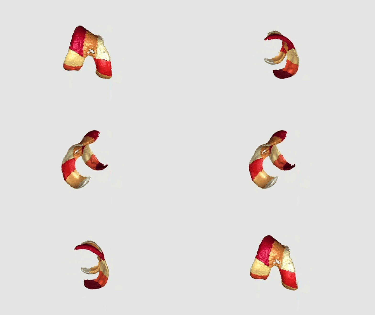
Chances are you have knee pain or know someone who does. Osteoarthritis, most commonly due to worn-down cartilage in the knee, affects over 230 million people worldwide, causing groans at best and an inability to live a full, mobile life at worst.
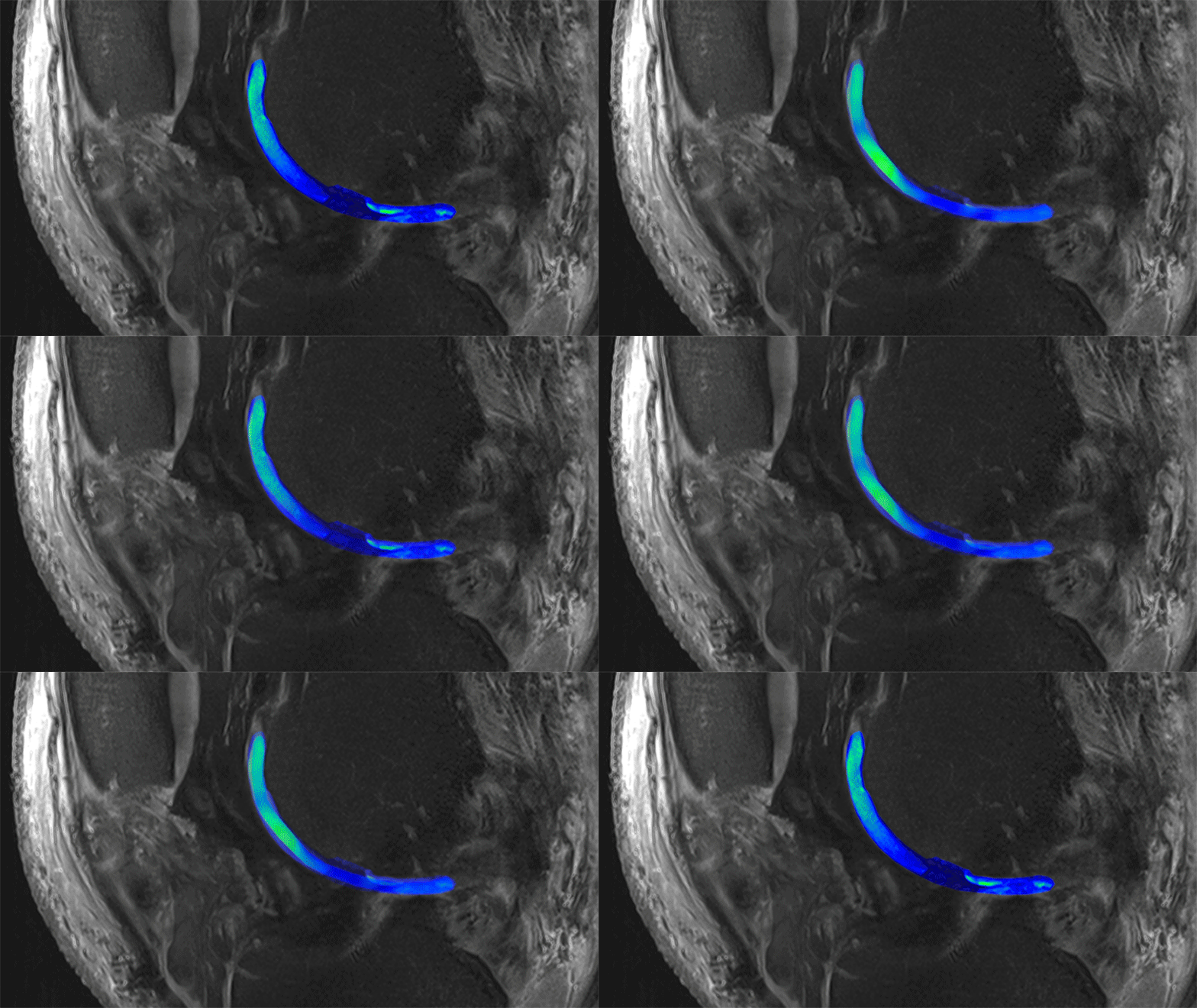
Novartis researchers are trying to regenerate functional cartilage in knees, but it’s hard to know if the experimental approach is working. Until now, the only way to find out has been to perform a biopsy to determine if the regenerated tissue is of high quality. To avoid the pain and injury of a biopsy, Novartis is collaborating with a scientist and his team at the Medical University of Vienna in Austria to measure the molecular makeup of regenerated cartilage using advanced magnetic resonance imaging (MRI) technology. This information could tell them if the regenerated tissue is the kind that will function like natural cartilage and help patients return to normal activities.
Their novel technique is giving them a way to see through knees.
Visualizing cartilage damage

What’s happening inside the knee to cause so much anguish? Underneath the kneecap lie several slips of tissue called cartilage.
Cartilage acts as a cushion that keeps bones from knocking together and has a smooth surface that enables joints to move freely. “The surface of cartilage is so slick, it’s like ice on ice,” says Didier Laurent, a Director in the Biomarker Development group at the Novartis Institutes for BioMedical Research (NIBR).
When osteoarthritis strikes, the cartilage breaks down. Pits form. That special springy and slippery function degrades. Joints become stiff and achy, and movement can become painful. The human body is not able to repair the damage.
Good cartilage defined

A protein called proteoglycan is part of what gives cartilage its spring. The material naturally weaves itself together with another protein, collagen, to create a super-strong material a superhero would envy. High-quality cartilage is resilient enough to withstand constant pounding and still spring back, and it is smooth enough to enable bones to move fast and free.
Since damaged cartilage does not regenerate on its own, Novartis scientists are working on a way to prod the body to regenerate these building blocks and weave them together into high-quality tissue. The challenge is to confirm that the regenerated cartilage is a powerful weave of collagen and proteoglycans, without, through a biopsy, having to cut out part of what they’ve just grown back.
A view into the knee

That’s where Siegfried Trattnig, an MRI expert at the Medical University of Vienna, and his MRI technology come in.
MRI machines in clinical use are typically 1.5 tesla or 3 tesla scanners (tesla is the measure of the magnetic field strength the machine produces). Trattnig, however, is working with a more powerful 7 tesla (7T) machine.
At 7T, it’s possible to see the finer details inside living bodies – such as knees – as small as 200 micrometers, slightly larger than the width of a human hair. The technology also generates information on tissue composition, which reflects how healthy the tissue is. This new capability is critical for picking up the subtle signals that reveal high-quality cartilage regeneration.


